


产品中心
- 细胞类
- 生化试剂
- ELISA检测
-
抗体蛋白
二抗生物素标记 过氧化物酶(HRP)标记 胶体金试剂 FITC荧光标记 RBITC荧光标记 二抗免疫血清 其它荧光标记二抗 藻红蛋白(PE)荧光标记 胶体金(Gold)标记 SAlexa Fluor荧光系列 碱性磷酸酶(AP)标记 别藻蓝蛋白(APC)荧光标记 其它标记 PE标记二抗 DyLight标记二抗 AU标记二抗 Biotin标记二抗 AMCA标记二抗 Texas Red标记二抗 TRITC标记二抗 HRP标记二抗 未标记二抗 Cy标记二抗 AbBox Fluor标记二抗内参抗体 小分子抗体抗体标记试剂盒细菌抗体蛋白病毒包装试剂杂交瘤融合筛选WB、IHC、ELISA相关试剂细胞培养试剂病原微生物抗原抗体假病毒抗体校准品其他抗原抗体标记的标签抗体病理级IHC抗体重组蛋白
- 细胞培养
- 实验耗材
- 仪器设备
- 生化试剂盒
- 小分子试剂
- 基质胶
-
斑马鱼产品
订货方式
订货电话:13564444959
订货时间:周一至周五
订货Q Q:79688691
订货邮件:79688691@qq.com
订货时间:周一至周五
订货Q Q:79688691
订货邮件:79688691@qq.com


产品详情
产品图片
注意事项
 产品详情
产品详情-
产品货号IM12055
-
别名GAPDH; GAPD; CDABP0047; OK/SW-cl.12; Glyceraldehyde-3-phosphate dehydrogenase; GAPDH; Peptidyl-cysteine S-nitrosylase GAPDH
-
产品名称GAPDH (2B8) Mouse Monoclonal Antibody
-
类别抗体产品
-
基因名称GAPDH
-
蛋白名称Glyceraldehyde-3-phosphate dehydrogenase
-
推荐应用WB,IF-ICC,IHC-P
-
反应种属human;Rat;Mouse;Mk;Dg;Ch;Hamster;Rabbit;Pig;sheep;Insect;Yeast;Bovine
-
存储缓冲液PBS, pH 7.4, containing 0.5%BSA, 0.02% New type preservative N as Preservative and 50% Glycerol.
-
Human Gene ID2597
-
Mouse Gene ID100042025
-
Rat Gene ID24383
-
免疫原Synthetic Peptide of GAPDH
-
特异性The antibody detects endogenous GAPDH protein.
-
稀释度WB 1:5000-20000, IHC-P 1:200-300, IF-ICC 1:200
-
参考分子量37kD
-
运输及保存条件-20°C/1 year
-
宿主Monoclonal, Mouse
-
背景介绍glyceraldehyde-3-phosphate dehydrogenase(GAPDH) Homo sapiens This gene encodes a member of the glyceraldehyde-3-phosphate dehydrogenase protein family. The encoded protein has been identified as a moonlighting protein based on its ability to perform mechanistically distinct functions. The product of this gene catalyzes an important energy-yielding step in carbohydrate metabolism, the reversible oxidative phosphorylation of glyceraldehyde-3-phosphate in the presence of inorganic phosphate and nicotinamide adenine dinucleotide (NAD). The encoded protein has additionally been identified to have uracil DNA glycosylase activity in the nucleus. Also, this protein contains a peptide that has antimicrobial activity against E. coli, P. aeruginosa, and C. albicans. Studies of a similar protein in mouse have assigned a variety of additional functions including nitrosylation of nuclear proteins, the regulation of mRNA stability, and acting as a transferri
-
纯化The antibody was affinity-purified from mouse ascites by affinity-chromatography using specific immunogen.
产品图片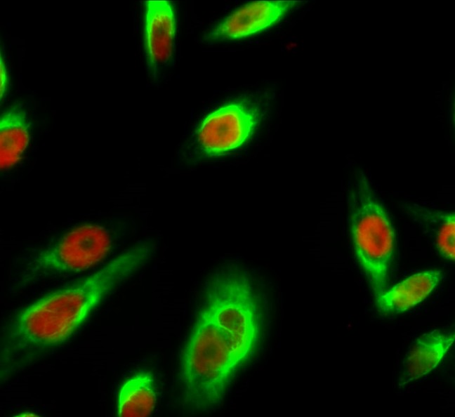 | Immunofluorescence analysis of Hela cell. 1,Cyclin D1 Polyclonal Antibody(red) was diluted at 1:200(4° overnight). GAPDH Monoclonal Antibody(2B8)(green) was diluted at 1:200(4° overnight). 2, Goat Anti Rabbit Alexa Fluor 594 was diluted at 1:1000(room temperature, 50min). Goat Anti Mouse Alexa Fluor 488 was diluted at 1:1000(room temperature, 50min). |
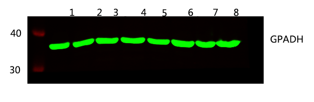 | Western blot analysis of 1 HEK293 2 SW480 3 HEPG2 4 MCF-7 5 mouse brain 6 Rat brain 7 Hela 8 A549 lysates, primary antibody was diluted at 1:5000, 4° over night, secondary antibody was diluted at 1:10000, 37° 1hour. |
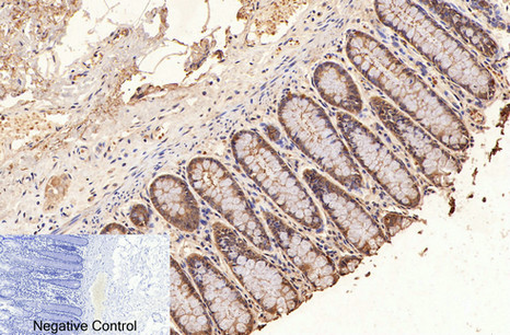 | Immunohistochemical analysis of paraffin-embedded Human-colon tissue. 1,GAPDH Monoclonal Antibody(2B8) was diluted at 1:200(4°C,overnight). 2, Sodium citrate pH 6.0 was used for antibody retrieval(>98°C,20min). 3,Secondary antibody was diluted at 1:200(room tempeRature, 30min). Negative control was used by secondary antibody only. |
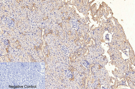 | Immunohistochemical analysis of paraffin-embedded Rat-kidney tissue. 1,GAPDH Monoclonal Antibody(2B8) was diluted at 1:200(4°C,overnight). 2, Sodium citrate pH 6.0 was used for antibody retrieval(>98°C,20min). 3,Secondary antibody was diluted at 1:200(room tempeRature, 30min). Negative control was used by secondary antibody only. |
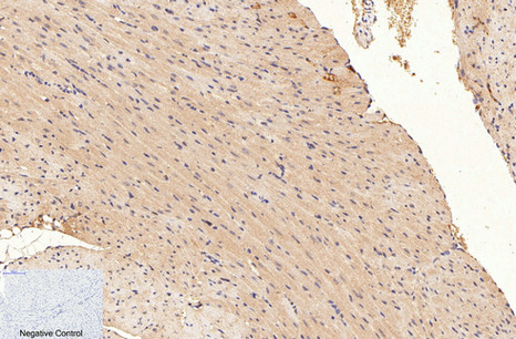 | Immunohistochemical analysis of paraffin-embedded Mouse-heart tissue. 1,GAPDH Monoclonal Antibody(2B8) was diluted at 1:200(4°C,overnight). 2, Sodium citrate pH 6.0 was used for antibody retrieval(>98°C,20min). 3,Secondary antibody was diluted at 1:200(room tempeRature, 30min). Negative control was used by secondary antibody only. |
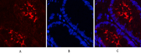 | Immunofluorescence analysis of Human-colon tissue. 1,GAPDH Monoclonal Antibody(2B8)(red) was diluted at 1:200(4°C,overnight). 2, Cy3 labled Secondary antibody was diluted at 1:300(room temperature, 50min).3, Picture B: DAPI(blue) 10min. Picture A:Target. Picture B: DAPI. Picture C: merge of A+B |
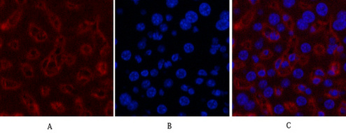 | Immunofluorescence analysis of Mouse-liver tissue. 1,GAPDH Monoclonal Antibody(2B8)(red) was diluted at 1:200(4°C,overnight). 2, Cy3 labled Secondary antibody was diluted at 1:300(room temperature, 50min).3, Picture B: DAPI(blue) 10min. Picture A:Target. Picture B: DAPI. Picture C: merge of A+B |
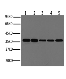
 | Western blot analysis of Hela (1), Rat brain (2), Rabbit Muscle (3), Sheep Muscle (4), and Mouse brain (5), diluted at 1:10000. |
注意事项1.本产品仅供科研使用。请勿用于医药、临床诊断或治疗,食品及化妆品等用途。请勿存放于普通住宅区。
2.为了您的安全和健康,请穿好实验服并佩戴一次性手套和口罩操作。
3.实验结果可由多种因素影响,相关处理只限于产品本身,不涉及其他赔偿。
备注:由于产品信息可能会有优化升级,请以实际收货标签信息为准。
产品推荐
-
DYKDDDDK-Tag(3B9) mAb¥500.00货号:IM72022
-
PCNA Monoclonal Antibody(12D10), HRP Conjugated¥2200.00货号:IM12505
-
Plant actin Monoclonal Antibody(Q30), HRP Conjugated¥2200.00货号:IM12506
-
Rubisco (Large Chain) Monoclonal Antibody(3G7), HRP Conjugated¥2200.00货号:IM12507
-
TBP/TATA Binding Protein Monoclonal Antibody(4H2),HRP Conjugated¥2200.00货号:IM12508